Leucoreduction: How White Cells Are Separated
By Lorna Tolentino, Flow Cytometry Laboratory Manager
As a blood donor, you likely already know that white blood cells are not transfused to patients. But do you know how and why the white cells are separated?
Leucoreduction is a filtration technique used to remove white blood cells, or leukocytes, from platelet and red blood cell products. White blood cells are known for the critical role they play in the body’s immune response, and part of the reason they can protect you so well is that white blood cells contain human leukocyte antigens. These are molecules on the white cell surface that are unique to each person and contain genetic information that codes how your body will respond to outside agents, among other things. And while these antigens are great for keeping you healthy, if your body were to be exposed to ones different than your own, there is a chance you could have a dangerous reaction.
For patients, being transfused with white cells could lead to febrile non-hemolytic transfusion reactions, HLA alloimmunization, CMV infections and/or transfusion-associated GVHD in immunocompromised recipients — all of which could severely endanger the patients’ lives.
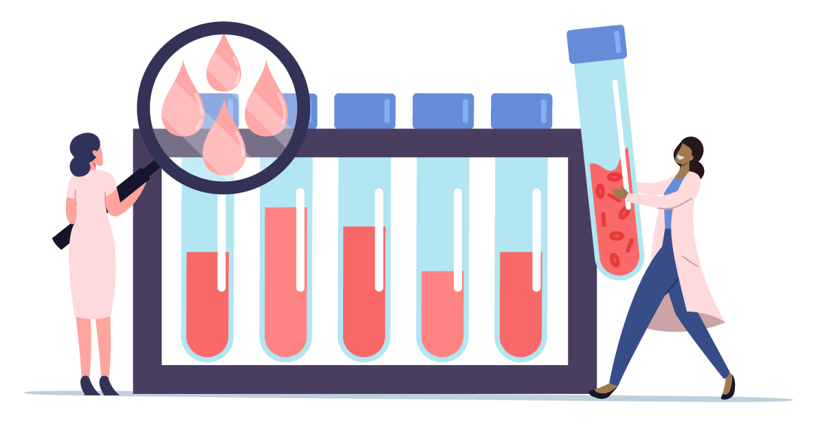
So, we’re agreed we do not want to transfuse donated white cells. Then how do we remove them? For platelets, white cells are removed automatically by the machine they’re being collected on; for whole blood, the standard method used by most blood centers is a fairly manual filtration process. Where it gets tricky for both of these methods, though, is quality control. We know that these methods are effective but, because of the great risk to patients, and because we know that no one system works 100% of the time, we need to check each product to make sure that the separation happened appropriately. To do this, laboratory team members historically would perform light microscopy, which involves looking under a microscope at a small sample of the product and actually counting how many white cell molecules are visible. While this methods is considered fairly accurate and is the most affordable option for blood centers, it’s also incredibly labor-intensive and tedious.
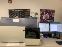
flow cytometer machine (left)
One of the benefits of having outstanding and innovate labs as part of our organization, however, is that SBC can utilize a newer, more efficient method: flow cytometry. With flow cytometry, the quality control happens without manual counting. Quality control team members manage this process by taking a small sample of the blood product, diluting it with a dye, and putting it into a tube with counting beads. This tube is then taken to the flow cytometry lab where it is placed in a flow cytometer machine, and the number of white blood cells remaining in the product is counted automatically, thereby obsoleting the labor-intensive process.
The most exciting part about this whole removal process is, of course, the multiple ways it supports patients. By separating out the white cells, not only do we minimize risk of transfusion-related reactions in patients, but also we can create special white cell products for researchers to use in conducting studies to improve the future of patient care!
So, on behalf of all of us at SBC, thank you for donating what you do — even the white cells! You’re saving more lives than you know.
Check out the rest of our Fall 2021 PULSE articles here!